|
R&D Systems
recombinant human galectin 3 protein  Recombinant Human Galectin 3 Protein, supplied by R&D Systems, used in various techniques. Bioz Stars score: 94/100, based on 1 PubMed citations. ZERO BIAS - scores, article reviews, protocol conditions and more https://www.bioz.com/result/recombinant human galectin 3 protein/product/R&D Systems Average 94 stars, based on 1 article reviews
recombinant human galectin 3 protein - by Bioz Stars,
2026-03
94/100 stars
|
Buy from Supplier |
|
R&D Systems
recombinant human gal 3 Recombinant Human Gal 3, supplied by R&D Systems, used in various techniques. Bioz Stars score: 93/100, based on 1 PubMed citations. ZERO BIAS - scores, article reviews, protocol conditions and more https://www.bioz.com/result/recombinant human gal 3/product/R&D Systems Average 93 stars, based on 1 article reviews
recombinant human gal 3 - by Bioz Stars,
2026-03
93/100 stars
|
Buy from Supplier |
|
OriGene
lgals3myc ddk Lgals3myc Ddk, supplied by OriGene, used in various techniques. Bioz Stars score: 90/100, based on 1 PubMed citations. ZERO BIAS - scores, article reviews, protocol conditions and more https://www.bioz.com/result/lgals3myc ddk/product/OriGene Average 90 stars, based on 1 article reviews
lgals3myc ddk - by Bioz Stars,
2026-03
90/100 stars
|
Buy from Supplier |
|
R&D Systems
gal 3 Gal 3, supplied by R&D Systems, used in various techniques. Bioz Stars score: 92/100, based on 1 PubMed citations. ZERO BIAS - scores, article reviews, protocol conditions and more https://www.bioz.com/result/gal 3/product/R&D Systems Average 92 stars, based on 1 article reviews
gal 3 - by Bioz Stars,
2026-03
92/100 stars
|
Buy from Supplier |
|
OriGene
human lgals3 protein  Human Lgals3 Protein, supplied by OriGene, used in various techniques. Bioz Stars score: 93/100, based on 1 PubMed citations. ZERO BIAS - scores, article reviews, protocol conditions and more https://www.bioz.com/result/human lgals3 protein/product/OriGene Average 93 stars, based on 1 article reviews
human lgals3 protein - by Bioz Stars,
2026-03
93/100 stars
|
Buy from Supplier |
|
Galectin Therapeutics
recombinant human galectin 3 protein (active) Recombinant Human Galectin 3 Protein (Active), supplied by Galectin Therapeutics, used in various techniques. Bioz Stars score: 90/100, based on 1 PubMed citations. ZERO BIAS - scores, article reviews, protocol conditions and more https://www.bioz.com/result/recombinant human galectin 3 protein (active)/product/Galectin Therapeutics Average 90 stars, based on 1 article reviews
recombinant human galectin 3 protein (active) - by Bioz Stars,
2026-03
90/100 stars
|
Buy from Supplier |
Image Search Results
Journal: bioRxiv
Article Title: Defining the mechanism of galectin-3-mediated TGF-β1 activation and its role in lung fibrosis
doi: 10.1101/2023.10.11.561855
Figure Lengend Snippet: Representative western blot of pSmad2 levels in (A) non-IPF HLFs (N=3) and (B) iHBECs (N=2) pre-treated with S0 μM SB-431S42 (ALKS inhibitor) or 1 μM GB0139 (galectin-3 inhibitor) for 20 minutes prior to 2-hour treatment with 10 μg/mL galectin-3 or 2 ng/mL TGF-β1. Western blot bands were quantified using densitometry analysis and presented as a ratio of pSmad2/tSmad2.
Article Snippet: After serum starvation, cells were stimulated with either 10 μg/mL
Techniques: Western Blot

Journal: bioRxiv
Article Title: Defining the mechanism of galectin-3-mediated TGF-β1 activation and its role in lung fibrosis
doi: 10.1101/2023.10.11.561855
Figure Lengend Snippet: Representative western blots of pSmad2 levels in non-IPF HLFs pre-treated with (A) NOTT199SS β1 inhibitor (0.1-100 nM) or (B-D) galectin-3 inhibitors GB0139, GB1107 and GB1211 (1 μM) or GB0149 (0.1-10 μM) for 20 minutes prior to stimulation with 2 ng/mL TGF-β1 (2-hour) or 50 μM LPA (4-hour). Cells pre-treated with S0 μM SB-431542 (ALK5 inhibitor) were included as a control demonstrating maximal inhibition of pSmad2 signaling. Western blot bands were quantified using densitometry analysis and presented as a ratio of pSmad2/tSmad2.
Article Snippet: After serum starvation, cells were stimulated with either 10 μg/mL
Techniques: Western Blot, Inhibition

Journal: bioRxiv
Article Title: Defining the mechanism of galectin-3-mediated TGF-β1 activation and its role in lung fibrosis
doi: 10.1101/2023.10.11.561855
Figure Lengend Snippet: Soluble galectin-3 (sequential injections, 19.5 - 5000 nM) binding to glycosylated or deglycosylated αv integrins: (A) αvβ1, (B) αvβS and (C) αvβ6 immobilised on the surface of a Series S sensor chip CM5 (approximately 1000 RU). (D) Soluble galectin-3 (sequential injections, 156.3-20000 nM) binding to glycosylated or deglycosylated TGFβRII immobilised to a Series S sensor chip CMS (approximately 400 RU). SPR signals were measured in RU and all sensorgrams baseline-corrected. Binding response values plotted in GraphPad Prism with connecting line/curve shown.
Article Snippet: After serum starvation, cells were stimulated with either 10 μg/mL
Techniques: Binding Assay
Journal: bioRxiv
Article Title: Defining the mechanism of galectin-3-mediated TGF-β1 activation and its role in lung fibrosis
doi: 10.1101/2023.10.11.561855
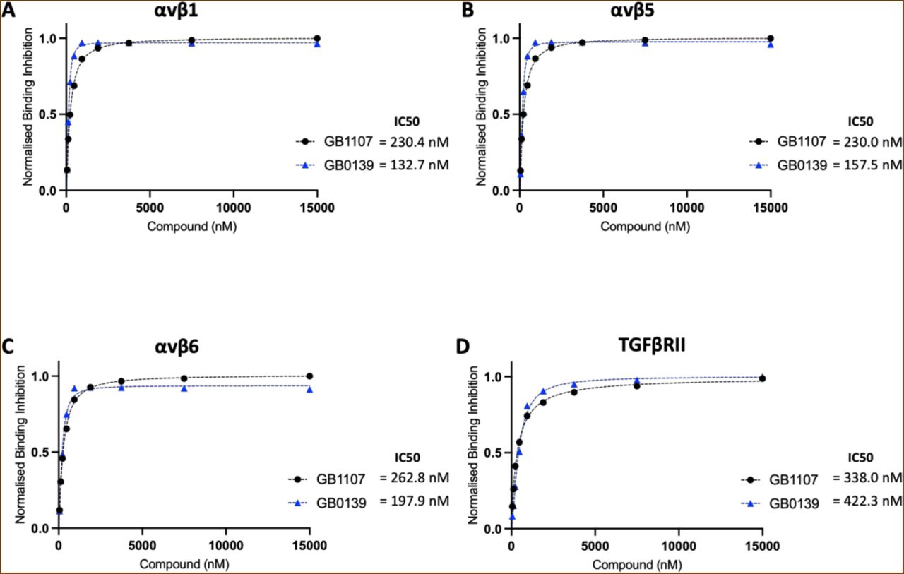
Figure Lengend Snippet: Solution competition binding assays performed with the galectin-3 inhibitor GB0139 (blue) or GB1107 (black) for αv integrins: (A) αvβ1, (B) αvβS and (C) αvβ6 or (D) TGFβRII in the presence of galectin-3 at 625 nM. Response values are normalised with respect to the highest binding response (DMSO control) and competitive inhibition graphs plotted in GraphPad Prism. IC50 values were calculated by non-linear regression analysis (binding saturation) - specific binding with hill slope.
Article Snippet: After serum starvation, cells were stimulated with either 10 μg/mL
Techniques: Binding Assay, Inhibition

Journal: bioRxiv
Article Title: Defining the mechanism of galectin-3-mediated TGF-β1 activation and its role in lung fibrosis
doi: 10.1101/2023.10.11.561855
Figure Lengend Snippet: Representative western blots showing co-immunoprecipitation of galectin-3 and the β1 integrin. Whole-cell protein lysates (6S0 μg/ IP reaction) from untreated non-IPF HLFs p6 (N=3) were immunoprecipitated with an anti-β1 integrin antibody (10 μg/ IP reaction) and immunoblotted for galectin-3 (upper panel) or immunoprecipitated with an anti-galectin-3 antibody (10 μg/ IP reaction) and immunoblotted for the β1 integrin (lower panel). Co-IP input, FT and wash steps loaded as controls. Proteins separated by reducing SDS-PAGE and target protein size estimated from the marker migration pattern.
Article Snippet: After serum starvation, cells were stimulated with either 10 μg/mL
Techniques: Western Blot, Immunoprecipitation, Co-Immunoprecipitation Assay, SDS Page, Marker, Migration

Journal: bioRxiv
Article Title: Defining the mechanism of galectin-3-mediated TGF-β1 activation and its role in lung fibrosis
doi: 10.1101/2023.10.11.561855
Figure Lengend Snippet: Representative confocal microscopy images (63x magnification) showing PLA of galectin-3 and the β1 integrin in (A) non-IPF HLFs p3-4 (N=3) or (B) IPF HLFs p3 (N=4) in the absence or presence of TGF-β1 stimulation (2 ng/mL TGF-β1 for 24 hours). Cells probed with a mouse anti-β1 integrin primary antibody (S μg/mL) and a rabbit anti-galectin-3 primary antibody (S μg/mL) followed by anti-rabbit PLUS and anti-mouse MINUS probes. Colocalization of galectin-3 and the β1 integrin 40 nm indicated by red fluorescence with DAPI counterstaining (blue).
Article Snippet: After serum starvation, cells were stimulated with either 10 μg/mL
Techniques: Confocal Microscopy, Fluorescence

Journal: bioRxiv
Article Title: Defining the mechanism of galectin-3-mediated TGF-β1 activation and its role in lung fibrosis
doi: 10.1101/2023.10.11.561855
Figure Lengend Snippet: (A) Downstream signaling of TGF-β1 following its integrin-mediated activation requires the integrin and TGF-β1 receptor to be in close proximity on the cell surface. (B) The galectin-3 carbohydrate binding domain binds to the glycosylation sites on αv integrins and the TGF-β1 receptor forming a galectin lattice at the cell surface which facilitates receptor clustering. This scaffold ensures that TGF-β1 can act on its receptor and potentiates TGF-β1 signaling. GB0139 binds to the galectin-3 carbohydrate recognition domain and blocks these protein-glycan interactions.
Article Snippet: After serum starvation, cells were stimulated with either 10 μg/mL
Techniques: Activation Assay, Binding Assay
Journal: Scientific Reports
Article Title: Targeting galectin-3 with a high-affinity antibody for inhibition of high-grade serous ovarian cancer and other MUC16/CA-125-expressing malignancies
doi: 10.1038/s41598-021-82686-3
Figure Lengend Snippet: Expression of LGALS3 and CA125 (MUC16) in human ovarian cancers. Three anonymized human tissue microarrays were constructed by collecting 1–3 cores from paraffin embedded, fixed blocks, as previously described . ( A ) Examples of human tumor cores stained for CA125 (panel i) and Gal3 (panel ii). Examples of core that did not stain for CA125 (panel iv) and tumor that did not stain for Gal3 (panel iii). ( B ) Each core was qualitatively scored for prevalence of CA125 and Gal3 positive cells (0 = < 5%, 1 + = 5–25%; 2 + = 25–50%; 3 + = 50–75% and 4 + = > 75%. The intensity for Gal3 was separately scored as low, medium and high (represented by green, orange and red dots respectively). All cores were independently scored by a blinded reference pathologist (KP).
Article Snippet: The last 2 immunizations used
Techniques: Expressing, Construct, Staining

Journal: Scientific Reports
Article Title: Targeting galectin-3 with a high-affinity antibody for inhibition of high-grade serous ovarian cancer and other MUC16/CA-125-expressing malignancies
doi: 10.1038/s41598-021-82686-3
Figure Lengend Snippet: Knockdown sh LGALS3- MDA-MB-231 cells show decreased invasiveness both in vitro and in vivo . ( a ) LGALS3 knockdown cell line, sh LGALS3 -MDA-MB-231 and a similar LGALS1 knockdown, sh LGALS1 -MDA-MB-231 were generated. LGALS3 and LGALS1 silencing was confirmed by Western blot in a manner previously described . β-Actin normalized densitometry quantification values are shown below each Western blot band. ( b ) In a Matrigel assay using triplicate chambers with 1 × 10 4 cells/chamber, invasion of the sh LGALS3 -MDA-MB-231 and sh LGALS1 -MDA-MB-231 were compared to the parental MDA-MB-231 wild-type cell line after 48 h ( p = 0.005, unpaired t test; error bars represent standard error). Three replicates were performed. ( c ) Representative luminescence images of all mice inoculated intravenously (by tail vein) with 5 × 10 6 sh LGALS3 -MDA-MB-231 cells or wild-type MDA-MB-231 control cells as previously described . ( d ) Kaplan–Meier survival curves; median survival for mice (n = 10 female athymic nude mice) implanted with MDA-MB-231 wild-type cells was 60 days (95% CI, 53.8–66.2), while median survival for mice (n = 10 female athymic nude mice) implanted with shLGALS3-MDA-MB-231 cells was not reached ( p = 0.02, log-rank test).
Article Snippet: The last 2 immunizations used
Techniques: In Vitro, In Vivo, Generated, Western Blot, Matrigel Assay

Journal: Scientific Reports
Article Title: Targeting galectin-3 with a high-affinity antibody for inhibition of high-grade serous ovarian cancer and other MUC16/CA-125-expressing malignancies
doi: 10.1038/s41598-021-82686-3
Figure Lengend Snippet: Anti-Gal3 monoclonal antibody, 14D11, inhibits MUC16-mediated tumorigenesis in vitro. ( a ) Direct ELISA for Gal3 binding to selected antibodies in the presence or absence of lactose. The presence of increasing lactose concentrations progressively inhibited the binding of Gal3 to 14D11, an isotype control antibody (18C6) and 1F5, an antibody reactive to the N terminal 15 amino acids of Gal3. Each lactose condition was compared to the zero lactose condition for the same antibody by paired t-test. ( b ) Effect of 14D11 on MUC16-mediated activation of ERK/AKT signaling in MUC16-expressing A2780 c344 and SKOV3 c344 cells after 48-h exposure to antibody. MUC16-negative A2780 and SKOV3 cells were used as controls. Densitometry was used to normalize the results against β-actin. ( c ) Effect of 14D11 on Matrigel invasion in MUC16-overexpressing SKOV3 c344 and A2780 c344 cells, and endogenous MUC16-expressing OVCAR3 cells. Anti-MUC16 N-glycosylation site antibody, 18C6, was used as a positive control. The values are the mean of triplicate well from a representative study and the invasion assays were repeated independently ≥ 3times. ( d ) Effect of 14D11 on Matrigel invasion in MDA-MB-231 and sh LGALS3 -MDA-MB-231 cells, performed as in 3C.
Article Snippet: The last 2 immunizations used
Techniques: In Vitro, Direct ELISA, Binding Assay, Activation Assay, Expressing, Positive Control